 |
| Tooth Development and Differentiation into Various Types https://www.ncbi.nlm.nih.gov/books /NBK27071/ |
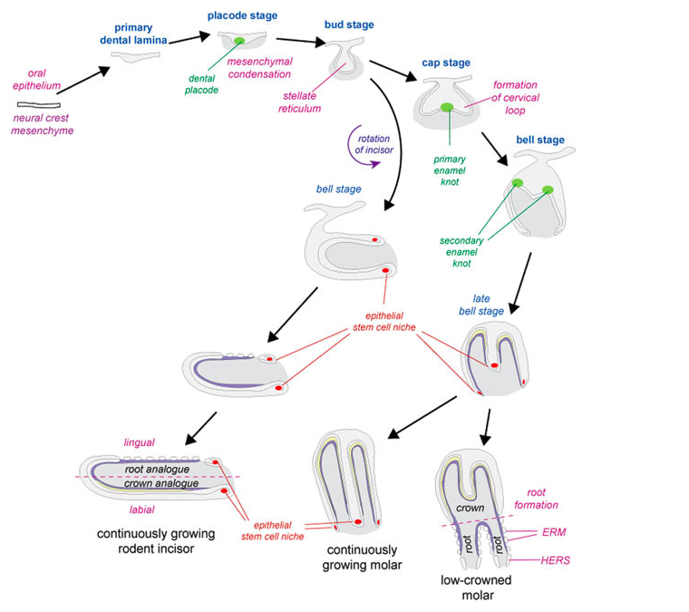
The hard tissues of the teeth - the enamel and dentin - are secreted by ameloblasts and odontoblasts that differentiate at the junction between the oral epithelium and the mesenchyme.
Following the development of the crown, root formation will occur and cementoblasts (differentiated from dental follicle mesenchyme) secrete cementum.
However, once teeth have matured most of the epithelial tissue has been lost (Thesleff et al., 2009). FUN FACT!!!: some rodents have continuously growing incisors due to epithelial stem cells that are maintained in the cervical loop. Since they are always gnawing on their food, this ensures that the incisors never wear down too far!
 |
| Dried, Non-Decalcified and Unstained Tooth Atlas of Histology |
 |
| Developing Tooth 1 Atlas of Histology |
These next two images show a tooth that is still developing. They show the rows of columnar ameloblasts and sections of enamel that contains calcified enamel rods. They also show the columnar odontoblasts that will secrete the
predentin, which can then calcify to form dentin
(Eroschenko et al., 2013).
 |
| Developing Tooth 2 Atlas of Histology |


No comments:
Post a Comment